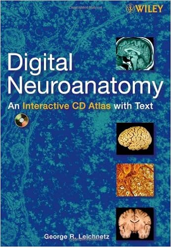
Download Digital Neuroanatomy by George R. Leichnetz PDF

By George R. Leichnetz
This multimedia source bargains an entire creation to neuroanatomy with fantastic, transparent and punctiliously classified photos and illustrations inside a sublime navigation constitution. It emphasizes the sensible points of the way to identify neuroanatomical constructions, with quizzes and bankruptcy self-assessments. The content material is organised into sections overlaying light-microscopic neurohistology, electron-microscopic neurohistology, skull-meninges-spinal wire, gross anatomy of the mind, sectional anatomy of the mind, and mind imaging.
Digital Neuroanatomy: An Interactive CD Atlas with evaluate Text features:
- Richly illustrated all through with over three hundred images
- A short revealed textbook that follows an analogous association and method, reviewing the entire major recommendations
- Self-grading quizzes with solutions that come with a close rationalization
- A aid mode providing lively reasons of the first programme positive factors
- A dynamic navigation constitution offering direct entry to express issues within the huge quantity of content material
An perfect device for instructing, self-instruction, and self-assessment, Digital Neuroanatomy: An Interactive CD Atlas with evaluation Text is a useful source for college students, teachers, and scientists alike. it truly is helpful for undergraduate classes and graduate classes in clinical, anatomy, radiology, dental, and pharmacy colleges, in addition to these in faculties of dentistry and actual remedy.
Content:
Chapter 1 Light?Microscopic (LM) Neurohistology (pages 1–19):
Chapter 2 Electron?Microscopic (EM) Neurohistology (pages 21–31):
Chapter three cranium, Meninges, and Spinal twine (pages 33–45):
Chapter four Gross Anatomy of the mind (pages 47–65):
Chapter five Sectional Anatomy of the mind (pages 67–76):
Chapter 6 advent to mind Imaging/MRIs (pages 77–85):
Read Online or Download Digital Neuroanatomy PDF
Similar anatomy books
Providing remarkable complete colour diagrams and scientific photographs, Langman's clinical Embryology, 13e is helping clinical, nursing, and well-being professions scholars strengthen a simple knowing of embryology and its scientific relevance. Concise bankruptcy summaries, fascinating scientific correlates containers, scientific difficulties, and a transparent, concise writing kind make the subject material available to scholars and correct to teachers.
Transduction Channels in Sensory Cells
This is often the 1st booklet to supply a molecular point clarification of the way the senses paintings, linking molecular biology with sensory body structure to infer the molecular mechanism of a key step in sensory sign new release. The editors have assembled professional authors from all fields of sensory body structure for an authoritative evaluation of the mechanisms of sensory sign transduction in either animals and vegetation.
Get Ready for A&P (Anatomy and Physiology)
Key gain: on hand as a workbook and site, this source saves school room time and frustration by way of helping readers speedy organize for his or her A&P direction. The hands-on workbook gets readers on top of things with simple examine abilities, math talents, anatomical terminology, uncomplicated chemistry, telephone biology, and different fundamentals of the human physique.
- Lung Mechanics: An Inverse Modeling Approach
- Color Atlas and Text of Histology
- The Determination of Molecular Structure
- Genome Editing: The Next Step in Gene Therapy
- An Atlas of Anatomy Basic to Radiology
Extra resources for Digital Neuroanatomy
Sample text
5b). INTERFASCICULAR OLIGODENDROCYTES In the CNS the white matter contains large numbers of myelinated axons that are easily identifiable due to the black density of the concentric lamellae of the myelin sheath surrounding the axons (Fig. 6a). Occasionally an EM section fortuitously shows an interfasicular oligodendrocyte, the neuroglial cell that produces the myelin sheath. Perineuronal oligodendrocytes are close to the cell body where they myelinate the first segment of the axon (Fig. 6b). SCHWANN-CELLS AND MYELINATED AND UNMYELINATED AXONS In the PNS the peripheral nerves contain Schwann cells (neurilemmal cells) whose membranous extensions wrap the axon in concentric lamellae of myelin (Fig.
2b EM dendritic neurotubules and neurofilaments. 3a EM dendrite with spine receiving axospinous terminal. Axon terminals or boutons contain abundant mitochondria and synaptic vesicles and typically end in synapses that show pre- and postsynaptic densities. Where these densities are heavier on the postsynaptic membrane, the synapse is asymmetric. Where the densities are roughly equivalent, the synapse is symmetric. 3b EM dendritic spine with spine apparatus and smooth ER. 4a EM axodendritic synapse with clear round synaptic vesicles.
Leichnetz Copyright # 2006 John Wiley & Sons, Inc. 1b Cerebral lobes. 2 Frontal lobe. Insular Lobe and Temporal Lobe 49 FRONTAL LOBE The precentral gyrus, the primary motor cortex, is the vertically running gyrus immediately in front of the central sulcus. It is the caudalmost gyrus of the frontal lobe. The remainder of the frontal lobe is made up of three gyri running horizontally perpendicular to the precentral gyrus, the superior, middle, and inferior frontal gyri separated by the superior and inferior frontal sulci (Fig.


