Download Digital neuroanatomy : an interactive CD atlas with text by George R. Leichnetz PDF

By George R. Leichnetz
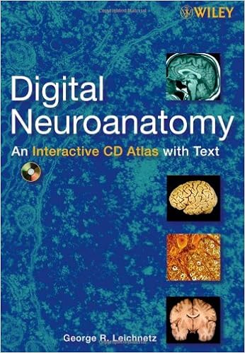
This multimedia source bargains an entire advent to neuroanatomy with remarkable, transparent and carefully categorised pictures and illustrations inside of a chic navigation constitution. It emphasizes the sensible facets of the way to identify neuroanatomical buildings, with quizzes and bankruptcy self-assessments. The content material is organised into sections overlaying light-microscopic neurohistology, electron-microscopic neurohistology, skull-meninges-spinal wire, gross anatomy of the mind, sectional anatomy of the mind, and mind imaging.
Digital Neuroanatomy: An Interactive CD Atlas with overview Text features:
- Richly illustrated all through with over three hundred images
- A short published textbook that follows an analogous association and strategy, reviewing the entire major concepts
- Self-grading quizzes with solutions that come with an in depth explanation
- A support mode providing lively motives of the first programme features
- A dynamic navigation constitution offering direct entry to precise issues within the huge quantity of content
An perfect instrument for educating, self-instruction, and self-assessment, Digital Neuroanatomy: An Interactive CD Atlas with overview Text is a useful source for college students, teachers, and scientists alike. it's priceless for undergraduate classes and graduate classes in clinical, anatomy, radiology, dental, and pharmacy colleges, in addition to these in colleges of dentistry and actual therapy.
Read Online or Download Digital neuroanatomy : an interactive CD atlas with text PDF
Similar anatomy books
Delivering unheard of complete colour diagrams and medical photos, Langman's scientific Embryology, 13e is helping scientific, nursing, and wellbeing and fitness professions scholars increase a simple realizing of embryology and its medical relevance. Concise bankruptcy summaries, alluring scientific correlates packing containers, scientific difficulties, and a transparent, concise writing kind make the subject material obtainable to scholars and suitable to teachers.
Transduction Channels in Sensory Cells
This can be the 1st ebook to supply a molecular point clarification of the way the senses paintings, linking molecular biology with sensory body structure to infer the molecular mechanism of a key step in sensory sign new release. The editors have assembled professional authors from all fields of sensory body structure for an authoritative evaluate of the mechanisms of sensory sign transduction in either animals and crops.
Get Ready for A&P (Anatomy and Physiology)
Key profit: on hand as a workbook and site, this source saves lecture room time and frustration by way of helping readers fast arrange for his or her A&P path. The hands-on workbook gets readers in control with easy research talents, math abilities, anatomical terminology, simple chemistry, phone biology, and different fundamentals of the human physique.
- The Cuvier-Geoffrey Debate: French Biology in the Decades before Darwin
- PARP Inhibitors for Cancer Therapy
- Myofascial and Fascial-Ligamentous Approaches in Osteopathic Manipulative Medicine
- Developmental Neurobiology
- Anatomy & Physiology Made Incredibly Easy!
Extra info for Digital neuroanatomy : an interactive CD atlas with text
Sample text
H & E. 11b Protoplasmic astrocyte and oligodendrocyte nuclei in cerebral cortex. Luxol fast blue/cresyl violet. PERIPHERAL NERVES: EPINEURIUM, PERINEURIUM, AND ENDONEURIUM In the PNS, peripheral nerves are covered with connective tissue. Epineurium is loose connective tissue and surrounds the entire nerve (Fig. a,b). The perineurium is denser connective tissue and surrounds individual fascicles of nerve fibers within the nerve. The endoneurium surrounds individual nerve fibers outside the myelin sheath.
The dorsal and ventral roots come together to form the spinal nerve at the level of the intervertebral foramen of exit. Spinal nerves C1 through C7 exit above the vertebra of the same number. C8 exits below vertebra C7. Then spinal nerves T1 through coccygeal-1 exit below the vertebrae of the same number. The spinal nerves in the cervical region exit nearly horizontally through intervertebral foramina at about the same level, but beginning in the thoracic region the dorsal and ventral roots descend to their foramen of exit.
11b Protoplasmic astrocyte and oligodendrocyte nuclei in cerebral cortex. Luxol fast blue/cresyl violet. PERIPHERAL NERVES: EPINEURIUM, PERINEURIUM, AND ENDONEURIUM In the PNS, peripheral nerves are covered with connective tissue. Epineurium is loose connective tissue and surrounds the entire nerve (Fig. a,b). The perineurium is denser connective tissue and surrounds individual fascicles of nerve fibers within the nerve. The endoneurium surrounds individual nerve fibers outside the myelin sheath.


