Download Human Brain Anatomy in Computerized Images by Hanna Damasio PDF
By Hanna Damasio
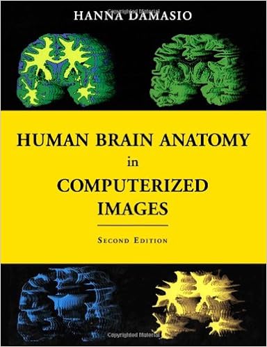
Sleek tomographic scans are revealing the constitution of the human mind in exceptional element. This spectator development, even though, poses a serious challenge for neuroscientists and practitioners of brain-related professions: how to define their means within the present tomographic photos with the intention to determine a specific mind web site, be it common or broken via ailment? the matter is made the entire tougher via the massive measure of person neuroanatomical edition. ready by means of a number one professional in complex brain-imaging ideas, this special atlas is a advisor to the localization of mind constructions that illustrates the wide variety of neuranatomical edition. it truly is in accordance with the research of 29 general mind acquired from 3-dimensional reconstructions of magnetic resonance scans of residing folks. It additionally presents 177 part (coronal, axial, and parasagital) of 1 of these brains in order that an identical constitution provided within the part got in a single occurrence will be pointed out within the component of one other occurrence. an extra 209 sections of 2 incidences of 2 different brains with diverse total configurations are integrated on the similar incidences, in order that readers can familiarize yourself with the range of normal photographs caused by way of assorted cranium shapes. Forty-six basic brains, segmented in to the main lobes, also are integrated. The atlas relies on a voxel-rendering process constructed within the author's laboratory that enables the reconstruction of the mind in 3 dimensions. The method allows the identity of significant sulci and gyri with in regards to the similar measure of precision that may be completed on the post-mortem desk. the quantity comprises 50 pages of colour illustrations. The Second Edition of this atlas deals completely new pictures, all from new mind specimens. just like the first variation, it's going to turn out to be a necessary software for neurologists, neurosurgeons, neuroradiologists, psychiatrists, and neuroscientists, in addition to clinical and neuroscience students.
Read Online or Download Human Brain Anatomy in Computerized Images PDF
Best anatomy books

Delivering unprecedented complete colour diagrams and scientific photos, Langman's scientific Embryology, 13e is helping scientific, nursing, and well-being professions scholars increase a simple figuring out of embryology and its scientific relevance. Concise bankruptcy summaries, eye-catching scientific correlates containers, scientific difficulties, and a transparent, concise writing sort make the subject material obtainable to scholars and appropriate to teachers.
Transduction Channels in Sensory Cells
This can be the 1st publication to supply a molecular point rationalization of ways the senses paintings, linking molecular biology with sensory body structure to infer the molecular mechanism of a key step in sensory sign iteration. The editors have assembled professional authors from all fields of sensory body structure for an authoritative evaluation of the mechanisms of sensory sign transduction in either animals and vegetation.

Get Ready for A&P (Anatomy and Physiology)
Key profit: on hand as a workbook and web site, this source saves school room time and frustration through helping readers fast organize for his or her A&P direction. The hands-on workbook gets readers up to the mark with uncomplicated learn abilities, math abilities, anatomical terminology, uncomplicated chemistry, mobile biology, and different fundamentals of the human physique.

- Computed Tomography of the Brain: Atlas of Normal Anatomy
- Drugs and Human Lactation
- Cardiac Gene Therapy: Methods and Protocols
- Advances in spinal fusion: molecular science, biomechanics, and clinical management
- Title Cognitive Foundations of Grammar
- Manual Mobilization of the Joints: Vol I The Extremities (6th Edition)
Additional info for Human Brain Anatomy in Computerized Images
Sample text
36] Figure 3-6 Bottom and top views (top] and front and back views (bottom) of Brachi-1 with marked gyri. See Chapter 2 for details. [37] Figure 3-7 Unroofing of the superior temporal gyrus. See Chapter 2 and legend of Figure 2-10 for details. [38] Chapter 4 Exterior Description of Another Brachicephalic Brain A nother brachicephalic brain, Brachi-2, is shown in this chapter. This is the JLJLbrain of a 34-year-old, fully right-handed man of Asian descent. The reason for depicting a second brachicephalic brain is that brachicephalic brains vary quite considerably among themselves.
It marks the mesial limit to the inferior temporal gyrus (ITG) or third temporal gyrus. The lateral limit of this gyrus is the inferior temporal sulcus. This gyrus is occupied by higher-order visual as- sociation cortex: Brodmann's field 20 anteriorly and the continuation of field 37 posteriorly. Normal Dolichocephalic Brain Mesial to the TOS, the most notable sulcus is the collateral (ColS). This sulcus marks the mesial limit of the fourth temporal gyrus, also known as the temporo-occipital gyrus (TOG) or fusiform gyrus of the temporal lobe.
20] Figure 2-4 Mesial views of Dolicho with marked gyri. See text for details. [21] Figure 2-5 Bottom and top views (top) and front and back views (bottom) of Dolicho with marked sulci. See text for details. [22] Figure 2-6 Bottom and top views (top) and front and back views (bottom) of Dolicho with marked gyri. See text for details. [23] Figure 2-7 Lateral views of Dolicho with Brodmann's cytoarchitectonic areas marked on the same views seen in Figure 2-2. See text for details. [24] Figure 2-8 Mesial views of Dolicho with Brodmann's cytoarchitectonic areas marked on the same views seen in Figure 2-4.