Download Human Sectional Anatomy: Atlas of Body Sections, CT and MRI by Harold Ellis, Bari M. Logan, Adrian Dixon PDF
By Harold Ellis, Bari M. Logan, Adrian Dixon
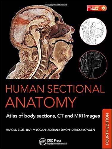
First released in 1991, Human Sectional Anatomy set new criteria for the standard of cadaver sections and accompanying radiological photographs. Now in its 3rd variation, this unsurpassed caliber is still and is extra stronger by means of a few worthy new fabric. As with the former versions, the wonderful full-color cadaver sections are in comparison with CT and MRI photos, with accompanying, labelled line diagrams. the various radiological photographs were changed with new examples, taken at the latest gear to make sure first-class visualization of the anatomy. thoroughly new web page spreads were further to enhance the book's insurance, together with photographs taken utilizing multidetector CT expertise, and a few appealing 3D quantity rendered CT photographs. The photographic fabric is more suitable via valuable notes, prolonged for the 3rd version, with info of vital anatomical and radiological positive aspects.
Read or Download Human Sectional Anatomy: Atlas of Body Sections, CT and MRI Images PDF
Similar anatomy books
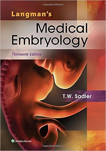
Delivering unheard of complete colour diagrams and scientific photos, Langman's clinical Embryology, 13e is helping clinical, nursing, and overall healthiness professions scholars strengthen a simple knowing of embryology and its medical relevance. Concise bankruptcy summaries, pleasing scientific correlates bins, medical difficulties, and a transparent, concise writing variety make the subject material obtainable to scholars and suitable to teachers.
Transduction Channels in Sensory Cells
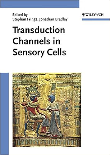
This can be the 1st publication to supply a molecular point rationalization of ways the senses paintings, linking molecular biology with sensory body structure to infer the molecular mechanism of a key step in sensory sign new release. The editors have assembled professional authors from all fields of sensory body structure for an authoritative evaluation of the mechanisms of sensory sign transduction in either animals and crops.
Get Ready for A&P (Anatomy and Physiology)
Key profit: on hand as a workbook and web site, this source saves school room time and frustration through helping readers fast arrange for his or her A&P direction. The hands-on workbook gets readers on top of things with easy examine talents, math abilities, anatomical terminology, easy chemistry, cellphone biology, and different fundamentals of the human physique.

- Annual Plant Reviews Volume 44: The Plant Hormone Ethylene
- Human Anatomy: Color Atlas and Textbook
- Neuroanatomy for the Neuroscientist
- A.D.A.M. Interactive Anatomy Online Student Lab Activity Guide
- A Laboratory Guide to Frog Anatomy
- Satureja: Ethnomedicine, Phytochemical Diversity and Pharmacological Activities
Additional resources for Human Sectional Anatomy: Atlas of Body Sections, CT and MRI Images
Sample text
The corona radiata (15) comprises a fan-shaped arrangement of afferent and efferent projection fibres, which join the grey matter to lower centres. On the computed tomography (CT) image, it appears as a curved linear area of low attenuation termed the centrum semiovale. The superficial temporal artery, of which the parietal branch can be seen at (7), is the smaller terminal branch of the external carotid artery, the other being the maxillary artery. The middle terminal branch can be seen immediately in front of (4).
The fourth ventricle (32) lies above the tegmentum of the pons (35) and below the vermis of the cerebellum (31). The ethmoidal air cells, or sinuses (6), are made up of some eight to ten loculi suspended from the outer extremity of the cribriform plate of the ethmoid bone and bounded laterally by its orbital plate. They thus occupy the upper lateral wall of the nasal cavity. The cells are divided into anterior, middle and posterior groups by bony septa. The middle group bulge into the middle meatus to form an elevation, the bulla ethmoidalis, into which they open.
Most of the intracranial venous sinuses are formed as clefts between these two layers, as demonstrated in this section by the superior sagittal sinus (6). The exceptions to this rule are the inferior sagittal sinus and the straight sinus, which are clefts within the meningeal layer. View ■ Orientation Anterior Right Left Posterior 6 2 6 11 Axial computed tomogram (CT) HEAD Axial section 2 – Male 1 6 7 8 9 2 5 4 6 1 2 3 4 5 6 7 8 9 12 3 Frontal bone Parietal bone Sagittal suture Dura mater Arachnoid mater Superior sagittal sinus Falx cerebri Subarachnoid space Pia mater Axial section 2 – Male ■ Section level HEAD ■ Notes This section, at a deeper plane through the skull vault, demonstrates the falx cerebri (7), which is formed as a double fold of the inner, meningeal, layer of the dura mater (5) and which forms the dural septum between the cerebral hemispheres.