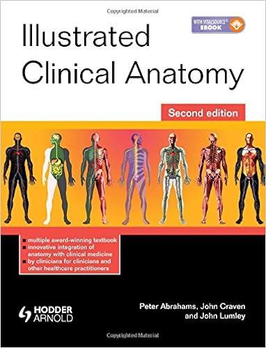
Download Illustrated Clinical Anatomy, Second Edition by Peter H. Abrahams, John L. Craven, John S.P. Lumley PDF
By Peter H. Abrahams, John L. Craven, John S.P. Lumley
Joint winner - Richard Asher Prize for the easiest new scientific textbook, RSM/Society of Authors booklet Awards 2005
Highly counseled within the 2005 BMA scientific booklet Awards: uncomplicated and medical Sciences category
"A outstanding e-book, very applicable, written with notable, didactic and shrewdpermanent illustrations! scientific anatomy at its best!" - Dr Ludmilla Stane, Institute for Anatomy, Dusseldorf.
The e-book of this new textbook in 2005 represented a big increase within the integration of anatomy educating with the research of medical medication. This re-creation builds in this well-received strategy, bringing the content material totally brand new with elevated emphasis at the stipulations most ordinarily encountered by way of scientific scholars and junior clinicians in training.
Structured through physique zone, every one bankruptcy comprises abundant scientific photos and photographs supplementing the fine quality anatomical diagrams, utilizing the easiest modality to illustrate anatomical relevance. Highlighted descriptions of medical relevance emphasise the built-in procedure so imperative to present instructing perform, and facilitated by way of the wealth of either medical and anatomical adventure of the prestigious writer team.
Read or Download Illustrated Clinical Anatomy, Second Edition PDF
Similar anatomy books
Supplying extraordinary complete colour diagrams and medical pictures, Langman's clinical Embryology, 13e is helping clinical, nursing, and healthiness professions scholars strengthen a easy realizing of embryology and its scientific relevance. Concise bankruptcy summaries, desirable scientific correlates bins, scientific difficulties, and a transparent, concise writing sort make the subject material obtainable to scholars and appropriate to teachers.
Transduction Channels in Sensory Cells
This can be the 1st publication to supply a molecular point rationalization of the way the senses paintings, linking molecular biology with sensory body structure to infer the molecular mechanism of a key step in sensory sign new release. The editors have assembled professional authors from all fields of sensory body structure for an authoritative review of the mechanisms of sensory sign transduction in either animals and vegetation.
Get Ready for A&P (Anatomy and Physiology)
Key gain: to be had as a workbook and site, this source saves lecture room time and frustration by means of helping readers quick arrange for his or her A&P path. The hands-on workbook gets readers up to the mark with uncomplicated learn abilities, math abilities, anatomical terminology, uncomplicated chemistry, telephone biology, and different fundamentals of the human physique.

- Developmental Neurobiology
- Anatomy: Embryology - Gross Anatomy - Neuroanatomy - Microanatomy
- Bodybuilding Anatomy-2nd Edition
- Eukaryotic Transcriptional and Post-Transcriptional Gene Expression Regulation
Additional info for Illustrated Clinical Anatomy, Second Edition
Example text
Bicuspid aortic valves are among the most common but are often symptom-free. A patent ductus arteriosus, atrial septal defects (caused by a persistent foramen ovale) and ventricular defects usually require surgical correction. If the ductus fails to close, pulmonary hypertension results, from the shunting of higher-pressure systemic blood into the pulmonary circulation, and cardiac failure usually follows. Surgical correction is required. Aortic coarctation is congenital narrowing of the aorta in the region of the ductus.
It flows to the rest of the body, but the majority passes via the umbilical arteries to the placenta. The venous return from the rest of the body, and the blood from the placenta in the umbilical vein and the ductus venosus, enters the inferior vena cava. At birth respiration begins and there is a consequent increase in blood flow to the lungs and an increase in venous return in the pulmonary veins. The left atrial pressure therefore rises and results in closure of the foramen ovale. The ductus venosus and ductus arteriosus also close at this time and, in doing so, establish the adult pattern of circulation.
B) Pericardial sac after removal of the heart. 18 Posterior aspect of the heart Aortic root Pulmonary trunk Right and left anterior cusps of the pulmonary valve. 19 Superior aspect of the heart showing site of origin of the great vessels and the pericardial reflections and sinuses pericardial sac rises. In these circumstances it is necessary to aspirate the fluid. The pericardial sac is most easily entered by a needle inserted between the xiphoid process and the left costal margin, directed headwards, backwards and medially through the central tendon of the diaphragm (Fig.