Download Serotonin Receptors in Neurobiology (Frontiers in by Amitabha Chattopadhyay PDF

By Amitabha Chattopadhyay
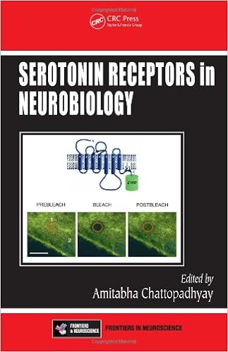
A few advancements spanning a mess of thoughts makes this a thrilling time for learn in serotonin receptors. A finished assessment of the topic from a multidisciplinary viewpoint, Serotonin Receptors in Neurobiology is one of the first books to incorporate details on serotonin receptor knockout reviews. With contributions from best specialists of their fields, the booklet explores serotonin receptors from a broad-based, multidisciplinary method. The ways defined fluctuate from molecular organic strategies to fluorescence microscopy and imaging, to genetic manipulation in animal versions, supplying quite a lot of instruments to check serotonergic phenomena. whereas each one of those techniques has its personal merits and barriers, the synthesis of data and data accomplished from reports utilizing a number of methods will lead to a complete figuring out of the underlying complicated phenomena curious about serotonergic signaling and its implications in healthiness and ailment. The e-book presents an total realizing of those receptors in keeping with at the moment used methodologies and methods. It describes particular experimental methods that may be of use to researchers drawn to addressing related difficulties concerning different G-protein-coupled receptor signaling structures.
Read or Download Serotonin Receptors in Neurobiology (Frontiers in Neuroscience) PDF
Best anatomy books
Providing unheard of complete colour diagrams and medical pictures, Langman's scientific Embryology, 13e is helping clinical, nursing, and wellbeing and fitness professions scholars improve a easy knowing of embryology and its medical relevance. Concise bankruptcy summaries, beautiful scientific correlates packing containers, medical difficulties, and a transparent, concise writing kind make the subject material obtainable to scholars and correct to teachers.
Transduction Channels in Sensory Cells
This is often the 1st e-book to supply a molecular point rationalization of ways the senses paintings, linking molecular biology with sensory body structure to infer the molecular mechanism of a key step in sensory sign iteration. The editors have assembled professional authors from all fields of sensory body structure for an authoritative evaluate of the mechanisms of sensory sign transduction in either animals and crops.
Get Ready for A&P (Anatomy and Physiology)
Key gain: on hand as a workbook and web site, this source saves school room time and frustration by means of helping readers quick organize for his or her A&P path. The hands-on workbook gets readers in control with uncomplicated learn abilities, math talents, anatomical terminology, simple chemistry, mobilephone biology, and different fundamentals of the human physique.
- Introduction to mathematical methods in bioinformatics
- Anatomy of the Cortex: Statistics and Geometry
- Wheat Diseases and Their Management
- Neuroanatomy in Clinical Context: An Atlas of Structures, Sections, Systems, and Syndromes
- Advanced Dietary Fibre Technology
Extra resources for Serotonin Receptors in Neurobiology (Frontiers in Neuroscience)
Example text
An area of interest was defined covering the cell and from the mean intensity of this area, the background intensity was subtracted. 6 (Color figure follows p. ) FRET analysis results from the experiments introduced in this chapter. CFP and YFP intensities were measured from a neuroblastoma cell N1E-115 transfected with Epac and 5-HT7 receptor. (A) Region of interest (blue) drawn around the cell as well as the background region (red) used for background correction. 12. 13 is shown as a green trace.
It will allow obtaining the FRET efficiency and the ratio between DA and D_A states in a quantitative manner. In contrast to many other techniques, this method is independent on the fluorophor concentration. Other methods to analyze FRET are fluorescence intensity based. Here the appearance of FRET will result in a measurable decrease of donor intensity and an increase of acceptor intensity. This property can be used to analyze FRET and to investigate the cAMP concentration. A common intensity-based method which is applied on confocal laser scanning microscopes (cLSM) is the acceptor photobleaching method.
The spatial resolution of the image is determined not only by the number of pixels on the chip but also by the optical resolution of the microscope. The intensity resolution, however, is determined by the bit depth of each pixel. The CCD chip in the iXon camera we used consisted of 512 × 512 pixels. Each pixel had a bit depth of 14 bit. Thus, signals are digitalized using up to 16,384 gray scale levels, which give a dynamic range of 84 dB. EXAMPLES In the experiments described next we used an Epac sensor and FRET analysis to examine effect of serotonin receptors on intracellular cAMP level.


