Download Visualization of Brain Functions: Proceedings of an by David Ottoson, etc. PDF
By David Ottoson, etc.
This e-book is a compilation of papers by way of scientists from numerous components of biophysical and biochemical options, with the purpose of exploring ways that multidiscipline methods could be dropped at deepen realizing of ways the mind works.
Read Online or Download Visualization of Brain Functions: Proceedings of an International Symposium held at The Wenner-Gren Center, Stockholm, 9–11 June, 1988 PDF
Best anatomy books
Providing unprecedented complete colour diagrams and scientific photos, Langman's scientific Embryology, 13e is helping scientific, nursing, and health and wellbeing professions scholars strengthen a easy figuring out of embryology and its scientific relevance. Concise bankruptcy summaries, attractive scientific correlates bins, medical difficulties, and a transparent, concise writing kind make the subject material obtainable to scholars and suitable to teachers.
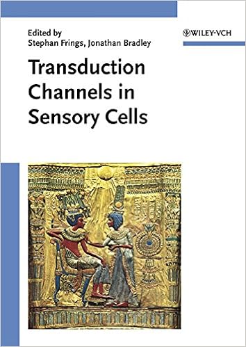
Transduction Channels in Sensory Cells
This is often the 1st publication to supply a molecular point rationalization of ways the senses paintings, linking molecular biology with sensory body structure to infer the molecular mechanism of a key step in sensory sign new release. The editors have assembled specialist authors from all fields of sensory body structure for an authoritative review of the mechanisms of sensory sign transduction in either animals and crops.
Get Ready for A&P (Anatomy and Physiology)
Key gain: to be had as a workbook and web site, this source saves school room time and frustration via helping readers fast arrange for his or her A&P path. The hands-on workbook gets readers in control with easy research abilities, math abilities, anatomical terminology, easy chemistry, cellphone biology, and different fundamentals of the human physique.
- Musculoskeletal Ultrasound: Anatomy and Technique
- PI3K-mTOR in Cancer and Cancer Therapy
- The Beagle Brain in Stereotaxic Coordinates
- The Proteins. Chemistry, Biological Activity, and Methods
- Schemata der Leitungsbahnen des Menschen
- Modeling the Optical and Visual Performance of the Human Eye (SPIE Press Press Monograph PM225)
Additional info for Visualization of Brain Functions: Proceedings of an International Symposium held at The Wenner-Gren Center, Stockholm, 9–11 June, 1988
Example text
De Vente and H. W. M. Steinbusch INTRODUCTION Chemical substances involved in the various aspects of neurotransmission are heterogeneously distributed. Besides specific intraneuronal fluorescence one has to reckon with extraneuronal fluorescen~e and non-specific fluorescent signals in the background. Consequently the application of quantitative i010unocytochemical methods to nervous tissues such as the brain encounters difficulties. It is not possible to put a measuring diaphragm around the substance to be investigated, as is feasible when the substance is located in a well defined morphological structure.
Indeed, for understanding of human chemical neuropathology, only human material can be used since most of neurological or psychiatric diseases especially degenerative and metabolic one are restricted to the human brain. II. ediately. aldehydesucrose-cryostat sections are used for neuropeptides using Sternberger's peroxidase antiperoxidase technique. Fresh cryostat sections are used for binding autoradiography or in situ hybridization. mediately used for radioiDDDunoassay or in vitro binding experillents.
In our laboratory summarized results obtained We will regarding, the distribution of several neuropeptides in the human hippocampal formation in adult as well as during development and aging, transient neuropeptide immunoreactivity in several parts of the developing brain and chemical neuropathology of a peculiar case of striatal atrophy. III. ANPAL I'OIII&TION and neuropeptide Y (NPY)(9) containing Interneurone somatostatine (SRIF)(B) are mainly concentrated in efferent parts of the hippocampal formation, in CAl, subicular complex and containing cholecystokinin Interneurone entorrhinal cortex.


